This website uses cookies so that we can provide you with the best user experience possible. Cookie information is stored in your browser and performs functions such as recognising you when you return to our website and helping our team to understand which sections of the website you find most interesting and useful.
Research
Seeing glaucoma from all angles
A new 3D eye model aims to give patients a clearer insight into their glaucoma and encourage them to stick with their treatment.
While current treatments for glaucoma are effective at stopping or slowing vision loss for many, a large number of people stop their treatments within several months of receiving their management plan.
Associate Professor Elaine Chong, CERA Senior Research Fellow and Head of Ophthalmology at the Royal Melbourne Hospital, says that arming patients with a better understanding of glaucoma is key to ensuring treatments are started early and taken regularly.
“When patients have a better understanding of what will happen if their condition deteriorates, they will be more motivated to take control of their treatment,” says Associate Professor Chong.
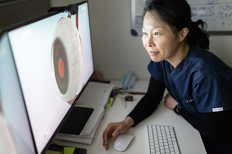
This led Associate Professor Chong and ophthalmology registrar Dr Joos Meyer to work with a team of designers and artists to develop One Right Eye: a 3D eye model quite literally modelled on a person’s right eye.
“In One Right Eye, you can actually see the damage caused to the optic nerve in glaucoma, which would otherwise be extremely hard to understand,” she says.
She says the model will also be a better way of working for ophthalmologists “because a picture tells 1000 words.”

How it works
Associate Professor Chong says explaining a complex eye condition like glaucoma to a patient without a visual aid is extremely difficult – especially during a short consultation.
“In glaucoma, nerves become sick and eventually die, and you need to be able to visually demonstrate to patients how it happens and why you need to monitor it.”
While physical eye models exist in some clinics, they are quite small and parts are often missing.
Associate Professor Chong says people with visual problems can look at the 3D model on an iPad or a computer in a lot more detail.
“It’s easier to understand because you can rotate the eye to see it from different angles and split the eye to see inside and zoom into a larger format.”
Increasing understanding
The software also allows clinicians to visually demonstrate the potential damage caused by cataracts, diabetic retinopathy and age-related macular degeneration.
The clinician selects the relevant condition in the program and then, using the 3D eye model, they can zoom in and out of the affected area to give the patient a visual understanding of the disease’s potential effects.
The team is currently running a trial of One Right Eye in clinics to assess patient satisfaction.
“Most patients really appreciate having a visual explanation of their condition rather than just a discussion,” says Associate Professor Chong.
The team is using feedback to refine the model and add features that can further improve patients’ understanding of eye disease.
New features will allow patients to see what happens during procedures such as visual field tests and OCT scans that check for eye diseases like glaucoma.
“For example, the patient will be able to see where an OCT scan is actually scanning on the eye – and how it relates to their condition,” says Dr Meyer.
Another feature in development will show patients a 3D view of how their vision may change and deteriorate over time.
After further refinements, the team hopes to roll out One Right Eye more widely into clinics.
This project is supported by CERA’s Innovation Fund and a Telematics Trust grant.

