This website uses cookies so that we can provide you with the best user experience possible. Cookie information is stored in your browser and performs functions such as recognising you when you return to our website and helping our team to understand which sections of the website you find most interesting and useful.
Research
Predicting glaucoma progression
The combined power of imaging and AI could identify people at risk of losing vision from glaucoma quickly – and who may benefit from new treatments.
Glaucoma treatments such as eye drops, laser treatment and surgery can help preserve many people’s sight, yet around one in three people still become blind in at least one eye within 20 years of being diagnosed with the condition.
“Predicting who will not respond well to routine treatment is one of the biggest challenges in managing glaucoma,” says Associate Professor Zhichao Wu, Head of Clinical Biomarkers Research at CERA.
Many new glaucoma treatments are being developed that aim to prevent damage in the eye before it occurs.
When these treatments eventually become available, clinicians will need better ways of identifying who is most at risk of losing vision quickly, as well as better methods of monitoring the effectiveness of these treatments.
Associate Professor Wu is working with CERA’s Ophthalmic Neuroscience team to use advanced imaging techniques in conjunction with AI to find signs – known as biomarkers – of cells that are sick and most likely to degenerate.
Identifying high-risk patients
Biomarkers are indicators of our body’s different processes, which can be used for diagnosing conditions, predicting worsening of disease and assessing the effectiveness of treatments.
Associate Professor Wu says there are not yet any known biomarkers that can accurately predict glaucoma progression.
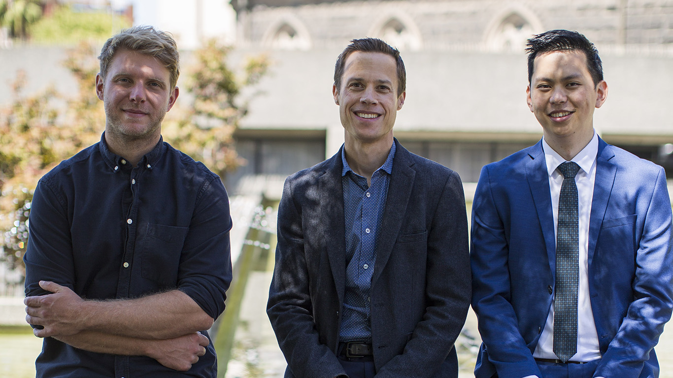
In the first part of the study, Associate Professor Wu, alongside Professor Peter van Wijngaarden and Dr Xavier Hadoux from the Ophthalmic Neuroscience Unit, is focusing on the potential of hyperspectral imaging.
A hyperspectral camera, developed at CERA, takes images of the retina with a multicoloured flash, enabling the team to see how the cells reflect light and find previously unseen signs of injury.
“The objective is detecting cells that appear sick, as these sick cells are usually the ones that will go on to die in glaucoma, causing progressive loss of vision,” says Associate Professor Wu.
“And that might give us a biomarker of the prediction for progression.”

Tracking glaucoma’s progress
With the use of AI, Associate Professor Wu says they aim to eventually produce a two‑dimensional picture of the back of the eye.
“We’re hoping for a heatmap of these sick cells to give an idea of how at risk patients are of progressing and where that damage will occur.
“Because not all damage is equal – damage closer to the center of your vision matters more for our daily activities.”
Once these high-risk patients are identified, clinicians will need a way of closely monitoring the effectiveness of their treatments.
However, the current method of tracking glaucoma progression can take years to pick up any vision loss.
“On average, it takes six years of doing visual field tests every six months to detect even moderate rates of visual field progression – and that’s shocking,” says Associate Professor Wu.
“In the second part of our study, we aim to compress this timeframe from six years to six months by picking up that change much sooner.”
Associate Professor Wu is using widefield optical coherence tomography (OCT) imaging combined with AI techniques, informed by knowledge of human anatomy and glaucoma to better distinguish between disease progression and healthy ageing.
“With this method, we aim to do a much better job of detecting glaucoma progression than is possible with visual field tests.”
This important study is following people with glaucoma over two years and is close to its halfway mark.
Associate Professor Wu and his team sincerely appreciate the commitment of research participants.
“We’re very thankful to the people that give up their time to help us gain insights into this disease.”

